Bladder Stones
BLADDER STONES
What are bladder stones?

Bladder stones, or more correctly called uroliths, are stones made up of minerals that form in the urinary bladder. They may occur as a large single stone or as collections of smaller stones.
How do bladder stones form?
There is a combination of reasons why bladder stones can form. They tend to form when there is increased levels of stone forming cystalline compounds in the urine. Diet as well as previous disease in the bladder can alter and affect this. When there is an increased level of cystalline compounds beyond what can be dissolved in the urine, it will preciptate and form crystals. With increased crystals in the urine, they can stick together and gradually form stones. Over time, these stones can increase in size and number.
What problems do bladder stones cause?
The two most common signs of bladder stones are
- Haematuria (blood in the urine): Blood in the urine occurs because the stones irritate the bladder wall, causing bleeding from its surface.
- Dysuria (straining to urinate): Straining to urinate occurs when stones obstruct the passage of urine out of the bladder.
When does it become an emergency?
When a stone gets lodged in the urinary tract, it may cause an obstruction. Obstructions are painful and can be life threatening due to a potential bladder rupture.
In these cases, patients may show the following signs:
- Profound weakness
- Haematuria (blood in the urine)
- Dysuria (Straining to urinate)
- Pollakiuria (Abnormal increase in urination frequency)
- Vomiting
- Distended and painful abdomen
When a full obstruction is present, the patient’s condition becomes critical and immediate therapy has to be done to relieve the obstruction. In certain cases, a partial obstruction may occur. This is not directly life threatening, however, it is important to note that it may develop into a full obstruction.
How are they diagnosed?
It is important to note that other bladder diseases may show similar signs as bladder stones (ie. Blood in the urine and/or straining to urinate). Further investigation are usually required to evaluate whether a patient does indeed have bladder stones.
Abdominal palpation may detect bladder stones or thickened bladder walls. However radiographs would give a better indication of how many stones and how big those stones are. The more common stones like struvite and calcium oxalate are able to be visualised on radiographs. Uncommon stones like urate and xanthine are not visible on radiographs and may require an ultrasound.
Ultrasonography can evaluate the size and thickness of the bladder wall. It can also confirm the presence and absence of stones, if any.
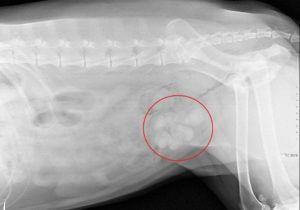

Figure 1 & 2: Bladder Stones seen in radiographs
How are bladder stones treated?
1. Cystotomy (Surgical Removal of Bladder Stones)
Surgical removal provides the fastest treatment option and in critical conditions, may be the only option. Surgery involves enterting the abdomen and bladder to manually remove the stones. However, this is limited to patients who have no secondary health conditions that can increase the risk of general anaesthesia.
2. Dissolution Diets
Some stones may be dissolved using special diets. This will avoid surgery and hence, be a better option for high risk patients. However, dissolution does not work for all types of stones. Moreover, the process is slow and can take weeks to months. Being a prescription diet, patients have to consume it exclusively, and this may not be the most palatable choice.
Please do not hesitate to contact us at 6455 6880 if you have any queries.