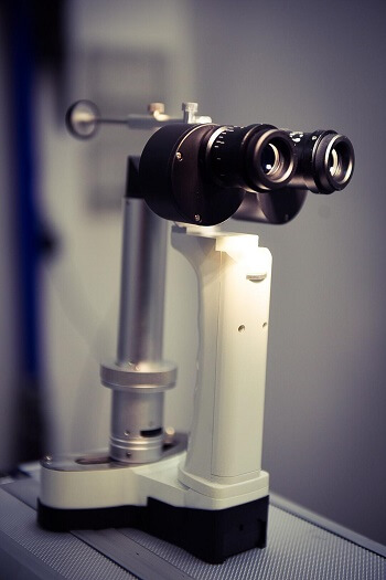
| What is a Slit Lamp?
A slit lamp is a tool that facilitates the examination of the front-half of an eye. The lamp consists of a high intensity variable light source and a set of binocular (stereoscopic) eyepieces. Adjusting the aperture of the light source creates a slit beam which allows the diagnosis of certain ocular conditions to be carried out. For a more detailed eye examination, the user can adjust the binocular eyepieces to increase its magnification. What are the diagnostic benefits of using a slit lamp? Distichiasis is a condition whereby fine hairs grow out of the glands in the eyelid margin and irritate the cornea surface. With the adjustable magnification of the slit lamp, such conditions can be picked up easily. Other examinations on the eye surface such as the curvature of various structures and abnormalities, indicating the presence of ulcer or masses, can also be detected by a slit lamp. |
 |
 |
A panoptic ophthalmoscope is a tool that allows us to examine the back of an eye and it is commonly used for retina examination. Evaluation of the cornea surface is also made possible with a special lens attached to it. When working in conjunction with a phone attachment, cornea and retina images can be easily captured and stored in the phone device for references. |
What are the diagnostic benefits of using it?
Studying the cornea surface and retina provides diagnostic information of hypertension, progressive retina atrophy and inflammation of optic nerve. With panoptic ophthalmoscope having a 5x higher magnification and a wider field of vision than a regular ophthalmoscope, it also allows the examiner to spot lesions easily.Fluorescein test is a technique specifically to assess the integrity of the cornea and detect the presence of any ulceration. Fluorescein is a green-tinted dye that will fluoresce under the blue light when the dye sticks on the exposed, injured area of the cornea.
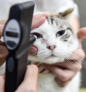
How is fluorescein test performed?A drop of fluorescein is applied to both eyes and both eyes are then examined under the blue light. A positive test will show green staining on the cornea at as the dye sticks on the injured area.
What are the diagnostic benefits of fluorescein test?A positive test will confirm the presence of corneal ulceration that needs to be addressed. Secondly, the size and color intensity of the dye will be the vet to assess the extent of the injuries and formulate appropriate treatment for the ulcers.
|
What is the Schirmer’s Tear Test?
Schirmer’s Tear Test is used to measure a patient’s tear production. The test must be performed without application of any topical medications or stains on the eye. Minimal restraint is required. What are the diagnostic benefits of the Schirmer’s Tear Test? Normal STT value is typically between 15 to 25mm per minute. STT is used to detect Dry Eye (Keratoconjunctivitis Sicca) |
 |
|
What is tonometry?
Tonometry is used to determine the intraocular pressure (IOP) of the eyes and is indicated for diagnosis of glaucoma where the IOP is elevated. How is tonometry performed?Tonometry is performed using a tonometer and will be done in a quiet environment to reduce stress level as much as possible. The tonometer will automatically calculate and average the IOP value of each eye individually. |
|
What are the diagnostic benefits of tonometry?
Tonometry is the main diagnostic tool for the diagnosis of glaucoma. It could be a primary issue or secondary to other ocular diseases. Further testing might be needed to find out these other causes of glaucoma and manage them accordingly.